Home » Without Label » Back Muscles Anatomy : Male Back Muscles Low Angle View Computer Illustration Anatomical 3d Stock Photo 308609242 / Together these muscles form a column, known as the erector spinae.
Back Muscles Anatomy : Male Back Muscles Low Angle View Computer Illustration Anatomical 3d Stock Photo 308609242 / Together these muscles form a column, known as the erector spinae.
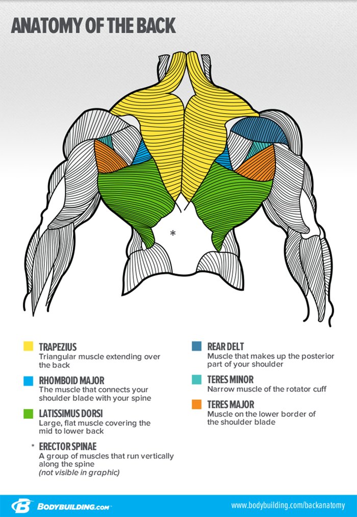
Back Muscles Anatomy : Male Back Muscles Low Angle View Computer Illustration Anatomical 3d Stock Photo 308609242 / Together these muscles form a column, known as the erector spinae.. All about the back muscles the back anatomy includes the latissimus dorsi, trapezius, erector spinae, rhomboid, and the teres major. To control the posture of the entire body. Leaning back to straight vertical and all points in between. These muscles give height and breadth to back development. This blog post article is an overview of the muscles of the lumbar spine of the trunk.
The back muscles are anatomically layered into superficial (extrinsic) and deep (intrinsic) muscles. To perform clinical clinical orthopedic manual therapy to the lumbar spine. The intrinsic back muscles are found deeper to the extrinsic muscles, separated from them by the thoracolumbar fascia. Superficial back muscles, intermediate back muscles and intrinsic back muscles.the intrinsic muscles are named as such because their embryological development begins in the back, oppose to the superficial and intermediate back muscles which develop elsewhere and are therefore classed as extrinsic muscles. These muscles provide posture and stability to the body by holding the vertebral column erect and adjusting the position of the body to maintain balance.
Lower Back Muscle Anatomy And Low Back Pain from marvel-b1-cdn.bc0a.com These muscles include the large paired muscles in the lower back, called erector spinae, which help hold up the spine, and gluteal muscles. The intrinsic back muscles are found deeper to the extrinsic muscles, separated from them by the thoracolumbar fascia. The back muscles are anatomically layered into superficial (extrinsic) and deep (intrinsic) muscles. The surface muscles of the upper back include the trapezius muscles (traps) and posterior deltoids. The deep muscles develop in the back called intrinsic muscles. The human spine is composed of 4 sections of vertebrae. The superior part of the appendicular skeleton that includes clavicle, scapula, and humerus, is attached to the axial skeleton that consists of skull. Muscle anatomy of the back 12 photos of the muscle anatomy of the back anatomy of the spine and.
The lower back (where most back pain occurs) includes the five vertebrae in the lumbar region and supports much of the weight of the upper body.
Ligaments hold the vertebrae in place, and tendons attach the muscles to the. Your lower back (lumbar spine) is the anatomic region between your lowest rib and the upper part of the buttock. Muscles of the lumbar spine. These are the muscles that are farther from the surface, closer to the internal organs and the spine. They start at the top of the neck and go down to the tailbone. See back muscle anatomy stock video clips. The extrinsic back muscles are located in the back, but act to produce movements of the shoulder and assist respiration. The muscles of the back can be arranged into 3 categories based on their location: Balance the weight of your head on top of your spine evenly distribute weights from your upper body into the lower extremities As a general group, they extend from the neck to the sacrum and fulfill a basic and fundamental function: Includes latissimus dorsi, the trapezius, levator scapulae and the rhomboids. These structures work together to support the body, enable a range of movements, and send messages from the brain to the. Muscle anatomy of the back 12 photos of the muscle anatomy of the back anatomy of the spine and.
Back pain is common and might be caused by a problem with a muscle. Superficial muscles of the back are located directly deep towards the skin along with superficial fascia.they are occasionally called the appendicular group as these muscles are mainly associated with activities of the appendicular skeleton. Related posts of back muscles chart muscle anatomy of the back. Three types of back muscles that help the spine function are extensors, flexors and obliques. This blog post article is an overview of the muscles of the lumbar spine of the trunk.
Back Anatomy All About The Back Muscles from www.kingofthegym.com What are the lower back muscles and their anatomy? All about the back muscles the back anatomy includes the latissimus dorsi, trapezius, erector spinae, rhomboid, and the teres major. The muscles of the back muscles make up a large part of the anatomy (structure) of the back. Deep muscles of the lower back include: As a general group, they extend from the neck to the sacrum and fulfill a basic and fundamental function: These structures work together to support the body, enable a range of movements, and send messages from the brain to the. They start at the top of the neck and go down to the tailbone. 1 your spine in this region has a natural inward curve.
The muscles of the back can be arranged into 3 categories based on their location:
On this page, you'll learn about each of these muscles, their locations and functional anatomy. The human spine is composed of 4 sections of vertebrae. The deep muscles develop in the back called intrinsic muscles. Extends and laterally bends the neck and head, rotates head to the same side: Related posts of back muscles chart muscle anatomy of the back. To control the posture of the entire body. All about the back muscles the back anatomy includes the latissimus dorsi, trapezius, erector spinae, rhomboid, and the teres major. The extrinsic back muscles are located in the back, but act to produce movements of the shoulder and assist respiration. Together these muscles form a column, known as the erector spinae. This curve, called lordosis, helps to: The extensor muscles are attached to back of the spine and enable standing and lifting objects. These are the muscles that are farther from the surface, closer to the internal organs and the spine. Understanding lower back anatomy is key to understanding the root of lower back and hip pain.
Mastoid process and lateral end of the superior nuchal line: Related posts of back muscles chart muscle anatomy of the back. On this page, you'll learn about each of these muscles, their locations and functional anatomy. Together these muscles form a column, known as the erector spinae. These sections are cervical (neck), thoracic (upper and middle back), lumbar (lower back), and sacrum (tailbone).
Your Blueprint For Building A Bigger Back from www.bodybuilding.com These muscles provide posture and stability to the body by holding the vertebral column erect and adjusting the position of the body to maintain balance. The spaces between the vertebrae are maintained by intervertebral discs that act like shock absorbers throughout the spinal column to cushion the bones as the body moves. The muscles of the lower back, including the erector spinae and quadratus lumborum muscles, contract to extend and laterally bend the vertebral column. See human back anatomy stock video clips. These muscles include the large paired muscles in the lower back, called erector spinae, which help hold up the spine, and gluteal muscles. Superficial back muscles, intermediate back muscles and intrinsic back muscles.the intrinsic muscles are named as such because their embryological development begins in the back, oppose to the superficial and intermediate back muscles which develop elsewhere and are therefore classed as extrinsic muscles. These are the muscles that are farther from the surface, closer to the internal organs and the spine. The deep muscles develop in the back called intrinsic muscles.
1 your spine in this region has a natural inward curve.
Muscles of the lumbar spine. These structures work together to support the body, enable a range of movements, and send messages from the brain to the. Leaning back to straight vertical and all points in between. This curve, called lordosis, helps to: To perform clinical clinical orthopedic manual therapy to the lumbar spine. The human spine is composed of 4 sections of vertebrae. Browse 3,558 back muscle anatomy stock photos and images available, or search for pelvic bone or lymphatic system to find more great stock photos and pictures. Back muscles the muscles of the back are a group of strong, paired muscles that lie on the posterior aspect of the trunk. These are the muscles that are farther from the surface, closer to the internal organs and the spine. The superior part of the appendicular skeleton that includes clavicle, scapula, and humerus, is attached to the axial skeleton that consists of skull. As a general group, they extend from the neck to the sacrum and fulfill a basic and fundamental function: The back muscles are anatomically layered into superficial (extrinsic) and deep (intrinsic) muscles. The deep muscles develop in the back called intrinsic muscles.